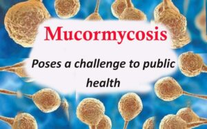
Mucormycosis in COVID- 19 patients in India poses a challenge to public health. Mucormycosis is caused by the fungi that belonged to the order Mucorales and class Zygomycetes. These fungi are widely prevalent in our environment.
COVID-19, an emerging life viral disease, is caused by a new severe acute respiratory syndrome (SARS-CoV-2) that was first time reported from China in 2019. The disease has rapidly resulted in a pandemic affecting over 187 countries of the world including India. Globally, more than 2.5 million people are infected with COVID-19 with a mortality rate ranging from 5 to 10%. In India, there are 2,96,33,105 cases and 3,79, 573 deaths from this devastating disease till June 16,2021. Covid-19 inflicted insuperable harm both to the human lives as well as global economy. It affects both sexes, and all age groups. However, elderly people with underlying medical conditions, such as diabetes mellitus, chronic obstructive pulmonary disease, cancer and chronic kidney disease are at a higher risk of getting COVID-19 infection. The maximum cases are observed in adults as compared to children.
Mucormycosis is an opportunistic life threatening mycosis that is reported from developed as well as developing nations of the world. The disease is caused by the fungi that belonged to the order Mucorales and class Zygomycetes. These fungi are widely prevalent in our environment. They are found in the soil, air, polluted water, decaying vegetables and fruits, stored grain, bread, rice, wheat, barley, groundnuts, dung, compost, etc. The fungi usually colonize the nose, sinuses, and eyes, and from there, it can reach to the brain. The causative agents of mucormycosis have preference for elastic lamina of large and small arteries causing hemorrhage, infarction and thrombosis. The disease can occur in sporadic and also epidemic form causing significant morbidity and mortality. Very recently, disease has posed another challenge in fighting the Covid menace.
Due to the dramatic surge of COVID-19 in India, the number of cases of mucormycosis has risen sharply posing a challenge to public health. It is reported that around 9,000 cases of mucormycosis have been diagnosed among COVID -19 patients from several states of India, such as Gujarat, Maharashtra, Madhya Pradesh, Karnataka, Rajasthan, Tamilnadu, Bihar, Odisha, Rajasthan, Telangana, Uttar Pradesh, Haryana, Chandigarh, and Delhi by May 22, 2021. It is pertinent to mention that maximum cases of mucormycosis are encountered in patients who were either suffering from chronic diabetics or undergone irrational therapy with corticosteroids.

Several clinical forms, such as cutaneous, subcutaneous, rhinocerebral, gastrointestinal, pulmonary, and systemic mucormycosis are recorded. Other unusual forms include pyelonephritis, endocarditis, and osteomyelitis. The disease has been diagnosed in patients with a history of uncontrolled diabetes, overuse of corticosteroids, malignancy, leukemia, extensive use of antibiotics, prolonged stay in intensive care unit (ICU), organ transplant, and voriconazole therapy. The clinical manifestation of mycormycosis in patients show fever, headache, shortness of breath, chest pain, coughing, bloody vomits, eyelid edema, pain and redness around eyes and nose, blurred or double vision, protruding eyes , sudden loss of vision, nasal discharge, stuffy nose, poor smell, facial paresthesia, skin lesion, toothache, and altered mental status.
Rhino-orbital-cerebral zygomycosis is most often encountered in patients with diabetes mellitus, and is recognized as a very severe form of the disease that carries mortality rate of 30 to 70%. It is pertinent to mention that around 40% of the cases of mucormycosis are related to diabetes mellitus. Globally, disease due to Rhizopus oryzae affects over 10,000 persons each year. The pulmonary infection can spread to other organs, if timely antifungal drug is not given to the patient.
The x-ray, computer tomography, and magnetic resonance imaging are helpful to spot the lesions in the body of the patient. The isolation of the fungus from the clinical specimens in pure and luxuriant growth on mycological media (Sabouraud dextrose agar, Pal sunflower seed agar, and APRM (Anubha, Pratibha, Raj, Mahendra) agar, and its direct microscopic detection as broad, aseptate hyphae in the wet mount, KOH (potassium hydroxide) preparation, PHOL (Pal, Hasegawa, Ono, Lee) stain , Narayan stain , and Gram`s stain are still widely used as the golden standard of diagnosis. In addition, histopahtological, and molecular techniques are also employed to confirm the disease. There is need to undertake further research to elucidate the role of immunological methods for the diagnosis of disease.
A combination of surgical debridement of necrotizing tissue if possible and antifungal therapy along with management of underlying disease is important to save the life of the patient. Immediate surgical removal of debridement of infected and necrotic tissues and administration of liposomal amphotericin-B (3-5 mg/kg body weight) helps to reduce the severity of disease. Very recently, posaconazole, a broad spectrum antifungal drug, has shown encouraging results in the patients suffering from mucormycosis. As liposomal amphotericin-B and posaconazole very expensive drugs, further research should be conducted to develop low cost, safe, and effective drugs that can be widely used for the better management of mucormycois, particularly by poor resource nations of the world.
Currently, there is no vaccine available for mucormycosis. Therefore, certain measures, such as judicial use of steroids, and antibiotics by the physician, correcting the predisposing factors, especially managing the diabetes mellitus, proper use of face mask by immuno compromised persons when visiting heavily polluted environment, avoiding contact with dirty surfaces, proper hand hygiene, avoiding to live in poorly ventilated, wet, moist and humid houses, early recognition of disease, and rapid institution of appropriate antifungal antibiotics will certain help to mitigate the incidence of mucormycosis in COVID-19 affected patients.
It is advised that to have a team of multidisciplinary experts that include microbiologist, ENT specialist, ophthalmologist, maxillofacial surgeon, neurologist, dentist, biochemist, and radiologist for the better management of this life threatening opportunistic fungal disease.


Mahendra Pal
Narayan Consultancy on Veterinary Public Health and Microbiology
Aangan, Jagnath Ganesh Dairy Road
Anand-388001, Gujarat, India
(palmahendra2@gmail.com)


