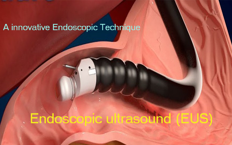
Endoscopic Ultrasound – a innovative Endoscopic Technique enables ultrasonographic images of high resolution to be obtained.Endoscopic ultrasonography is now a widely accepted modality for the diagnosis of pancreatobiliary diseases.
Endoscopic ultrasound (EUS) or echo-endoscopy is a medical procedure in which endoscopy (insertion of a probe into a hollow organ) is combined with ultrasound to obtain images of the internal organs in the chest, abdomen and colon. It can be used to visualize the walls of these organs, or to look at adjacent structures. Combined with Doppler imaging, nearby blood vessels can also be evaluated.
In this technique use of high frequency transducer, which, due to the short distance to the target lesion, enables ultrasonographic images of high resolution to be obtained. Endoscopic ultrasonography is now a widely accepted modality for the diagnosis of pancreatobiliary diseases. It can be used to determine the depth of invasion of gastrointestinal malignancies, and often for visualizing lesions more precisely than other imaging modalities.
Endoscopic ultrasound is performed with the patient sedated. The endoscope is passed through the mouth and advanced through the esophagus to the suspicious area. From various positions between the esophagus and duodenum, organs within and outside the gastrointestinal tract can be imaged to see if they are abnormal, and they can be biopsied by a process called fine needle aspiration. Organs such as the liver, pancreas, bile duct, gall bladder, adrenal glands, abnormal lymph nodes in chest and abdomen are easily biopsied.
In addition, the gastrointestinal wall itself can be imaged to see if it is abnormally thick, suggesting inflammation or malignancy. Advantage of tissue sampling by this technique is less painful, more accurate, minimal adverse efffects and without any chance of tumour spread while sampling. The technique is highly sensitive for detection of pancreatic cancer. It provides an excellent imaging modality for diagnosis and staging of pancreatic carcinoma.


EUS is now main stay of treatment for any fluid collection around pancreas which formed due to pancreatic inflammmation. Some time this modalitity use to diagnosis cause of pancreatic inflammation.
Bleeding from Gastric varices is most fearsome compliation may cause bloody vomiting can be life threating if bot controlled timely. Obliteration of gastric varices requires injection of occlusive agents (Cyanoacrylate glue and metallic coils) in bleeding gastric varices. EUS enables not only monitoring of occlusive agents during the procedure but also visualization of blood flow, the size of the varix to which coil deployment is planned, and obliteration of the vessel cavity following cyanoacrylate (CYA) injection. Therefore, it is quite advantageous to use EUS during this procedure to avoid risk of embolization of occlusive agents.


Dr.Kapil Sharma
Senior Consultant -Gastroenterology
Batra Hospital, New Delhi.


